
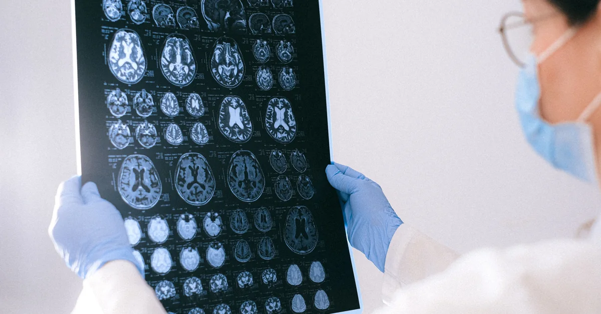
A large UK study finds that MRI measures of the choroid plexus—a structure that makes cerebrospinal fluid—track with later dementia risk. The finding is a signal, not a test.
Three things to remember
- 45,306 UK Biobank participants were followed over time.
- Larger choroid plexus and lower signal intensity raised risk.
- This is an association, not a diagnostic tool.
How to interpret it
Historical Biophysics & Lineage
The modern study uses MRI to measure choroid plexus volume and signal intensity, which are proxies for its secretory and barrier functions. Cserr’s work established that the choroid plexus produces CSF and regulates solute exchange, and that its dysfunction leads to impaired clearance of metabolic waste and altered brain fluid dynamics. The observed association between larger choroid plexus and lower signal intensity with increased dementia risk aligns with this: chronic inflammation, fibrosis, or calcification of the choroid plexus (reflected in MRI signal changes) can reduce CSF turnover, impair glymphatic clearance, and promote accumulation of amyloid and tau proteins. This mechanistic link validates Cserr’s hypothesis that choroid plexus integrity is critical for long-term brain health, and it extends her physiological findings to a clinical, population-level biomarker.
Source
This signal is based on Brain fluid structure may flag future dementia risk years before diagnosis from Medical Xpress Healthy Aging.
Primary study: Molecular Psychiatry — The choroid plexus, cognitive decline, and incident dementia.
Health note: This is an observational finding. It does not prove cause and effect, and it is not a clinical test. More research is needed.